Research Article
Cutaneous Larva Migrans in a Newborn of 3 Weeks
1344
Views & Citations344
Likes & Shares
Also referred to as creeping dermatitis, Cutaneous
Larva Migrans (CML) is a parasitic disease caused by hookworm larvae in a dead
end in moist soil. More common in children, it is rare in adults and
exceptional in infants. We report a case in a 3-week-old Newborn that was sent
to us by the pediatric department of the National Hospital of Niamey.
Contamination was made by contact with clothes soiled by moist soil containing
larvae. The presence of stray cats in the house was confirmed by the mother.
Local treatment with albendazole in preparation with a cream resulted in
complete healing after 2 weeks.
Keywords: Cutaneous
Larva Migrans, Newborn, Albendazole, Niger
INTRODUCTION
Cutaneous Larva Migrans (CML), also known as
"migrant helminthiasis", "dermatitis serpiginosus",
"creeping eruption", or "sand worm" is a zoodermatosis
caused by cutaneous penetration of helminth larvae, usually parasites of the
small bintestines of cats and dogs [1]. It is first described in 1874 by Lee RJ
[2], as a benign skin condition. More noticeable in tropical and subtropical
countries, it is caused by the accidental migration of an animal nematode larva
into the surface part of the skin. The most incriminated larvae are: Ancylostoma caninum, Ancylostoma brasiliense
and Uncinaria stenocephala [3].
We report a case in a 3-week-old Newborn that was sent to us by the pediatric
department of the National Hospital of Niamey. Local treatment with albendazole
as a mixture in a cream, led to complete cure after 2 weeks.
OBSERVATION
This was a three-week-old newborn that we received in consultation with the
Department of Dermatology at the National Hospital of Niamey, Niger (west
Africa). The duration of the illness before the consultation was 7 days. Crying
and agitation link to prurit was the reason for pediatric consultation. No
treatment has been done locally and by the general route. Physical examination
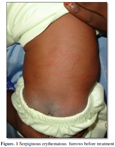
revealed numerous serpiginous erythematous lesions at the back without
excoriation, measuring several centimeters in length (Figure 1). At the interrogation, the mother of the baby revealed
the presence of stray cats in the family environment. After the laundry, the
newborn's clothes are spread out in the courtyard of the house on a rope where
some of them fall to be in contact with the soil soiled with larvae. The
general condition of the newborn was normal, without any other cutaneous
affection. Base line hematological and biochemical investigations were within
normal limits. According to epidemiological and clinic aspects, diagnosis of
CLM was made. A local treatment
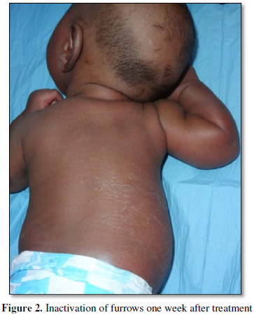
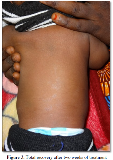
combining albendazole mixed in a moisturizing cream was carried out. One week
after initiation of treatment, inactivation of serpiginous erythematous furrows
(Figure 2) was observed; two weeks
later, the total disappearance of the furrows was seen (Figure 3). No local side effects were noted.
DISCUSSION
Rare in temperate countries, CML is reported mainly in tropical countries
[4,5]. The reported cases mainly concern the 1 to 5 year age group [6]; adults
appear to be more affected during travel due to the presence of cats roaming on
beaches and hotels [7-9]. The peculiarity of this observation is the appearance
of Cutaneous Larva Migrans in a newborn of three weeks; Classically, the
serpiginous erythematous lesion is objective [5,7,8,10] and was present in this
newborn. The location of the lesions is mainly in the areas most in contact
with the damp and soiled soil and concerns the pelvic limbs [7, 11,12]; in this
small patient, contamination of the back skin, was indirect through the drying
clothes that fell on a moist soil containing larvae. Many molecules used orally
alone or in combination have proved to be effective, the main ones being:
albendazole, thiabendazole, mebendazole, and / or ivermectin [13]. The local
treatment performed with thiabendazol [14] on another occasion also responds
well with albendazol, with which our patient was cured in 2 weeks without side
effects such as allergic contact dermatitis, pruritus and irritation [15].
CONCLUSION
The cutaneous larva migrans is classically favored by moisture and domestic
host animals mainly cat and dog.
Neonatal infestations are exceptional and
would be favored by wearing clothes soiled by wet soil containing larvae. Local
treatment based on albendazole in a cream is always effective.
STATEMENT OF INFORMED CONSENT
Written informed consent was obtained from the
patient’s father for publication of this article and any accompanying images.
CONFLICT OF INTEREST
None declared
- Meotti
CD, Plates G, Chagas Nogueira LL, da Silva RA, Paolini KS, Nunes EM et al.
(2014) Cutaneous larva migrans on the scalpatypical presentation of a
common disease. Ann Bras Dermatol 89: 332-333.
- Lee
RJ (1874) Case of creeping eruption. Trans Clin Soc London 8: 44 -45.
- Romain
B, Christian C, Tristan F (2016) Imported cutaneous larva migrans by a 31-
year-old French woman after a travel in Gabon. BMJ Case Rep.
- Tamminga
N, Bierman Wouter FW, De Vries PJ (2009) Cutaneous Larva Migrans acquired
in
- Brittany France. Emerg
infect Dis 15: 1856-1857.
- Camara A, Camara AD, Baldé H, Soumah MM, Keita M, Doumbouya A, et
al. (2011) Larva migrans cutanée: aspect épidémiologique, clinique et
thérapeutique. Ann Dermatol
Vénéréol 138s: 296.
- Salissou L, Adehossi E, Brah S, Gado M, MaguiaT, Kanga JM (2012)
Larva Migrans Cutanée: Aspect épidémiologique, Clinique, et thérapeutique
à propos de 73 cas au Centre National Dermato-lèpre de Niamey (Niger). Ann
Univers ABDOU M, Tome XIII-A, PP72-76.
- Jelineck
T, Maïwald H, Northdurft HD, Löscher T (1994) Cutaneous Larva Migrans in
travelers:synopsis of Histories, symptoms and treatements of 98 patients. Clin Infect Dis 19: 1062-1066.
- Caumes E, Carrière J, Guermonprez, G Bricaire F. et al. (1995) Dermatosis
Associated with Travel to Tropical Countries: A prospective study of the
diagnosis and management of 269 patients presenting to a tropical disease
Unit. Clin Infect Dis 20: 542-548.
- Bouchaud
O, Houzé S, Schiemann R, Durand R, Ralaimazaba P. et al. (2000) Cutaneous
larva migrans in travelers: A prospective study, with assesment of therapy
with ivermectin. Clin Infect Dis 31: 493-498.
- Davies HD, Sakuls P, Keystone JS (1993)
Creeping Eruption. A review of clinical presentation and management of 60
cases presenting to a tropical disease Unit. Arch Dermatol 129: 588-91.
- Prudhomme L, Loche F, Massip P, Marchou B (2002) Larva migrans
cutanée : Echec de l'ivermectine en dose unique. Méd Mal Infect 32:
115-118.
- Torres J, Orihuela A, Garcia D,
Abdul-Hadi S (1989) Treatment of cutaneous larva migrans with Albendazole.
Preliminary Report. Rev Inst Med 31: 56-58.
- Joseph
AM, Shafi KM, Das V, Soman S, Athira RS, Varghese T (2017) Cutaneous Larva
Migrans- A Case Study. World J Pharm 6: 1303-1307.
- Camparin
C, Rodrigues MM, Santos BC (2016) Extensive Cutaneous Larva Migrans with
Eczematous Reaction on atypical Localization. Ann J Trop Med Hyg 94:
1185-1186.
- Chabasse D, Le Clec’h C, de Gentile L, Verret JL (1995) Le larbish.
Cahier Santé 5 : 341-345.
QUICK LINKS
- SUBMIT MANUSCRIPT
- RECOMMEND THE JOURNAL
-
SUBSCRIBE FOR ALERTS
RELATED JOURNALS
- Stem Cell Research and Therapeutics (ISSN:2474-4646)
- Journal of Forensic Research and Criminal Investigation (ISSN: 2640-0846)
- International Journal of AIDS (ISSN: 2644-3023)
- Ophthalmology Clinics and Research (ISSN:2638-115X)
- Journal of Spine Diseases
- Journal of Alcoholism Clinical Research
- Journal of Clinical Trials and Research (ISSN:2637-7373)


